Can I obtain VA disability benefits for lung disease?
Yes, VA disability benefits for lung disease may be available.
You will need to prove that (a) you were in the military, (b) your lung disease originated or was aggravated while you were on active duty, (c) you were continuously treated for your lung disease since leaving the service (unless you are filing your disability claim within one year of leaving the service or your condition has been chronic), and (d) you are currently disabled by your lung disease.
About lung disease
Red blood cells must be brought as closely as possible to the air we breathe, so that hemoglobin in the cells can give up waste carbon dioxide from cellular metabolism and take on oxygen.
To accomplish this, the lungs have millions of tiny air sacs (alveoli) with very thin walls (alveolar membranes) containing microscopic blood vessels (capillaries). This anatomy of the lungs allows exposure of a large surface area of the blood to the air. Oxygen (O2) and carbon dioxide (CO2) gases diffuse (move) across the one cell thick alveolar membrane in opposite directions with the oxygen entering the blood and the carbon dioxide leaving it. This process is known as gas exchange.
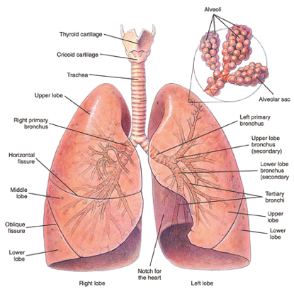
Figure 1: Bronchi and lungs.
The gas exchange part of the lungs is known as the lung parenchyma. Air is delivered to the parenchyma of the lungs through the bronchial tree — a repetitively branching tubular system for air conduction. It consists of the trachea, from which arises a right and left main (primary) bronchus to the right and left lungs. Smaller bronchi branch from the main bronchus of the right or left lung, then to smaller bronchi to the various lobes of the lungs, then to even smaller bronchi (bronchioles) that eventually reach the alveoli.
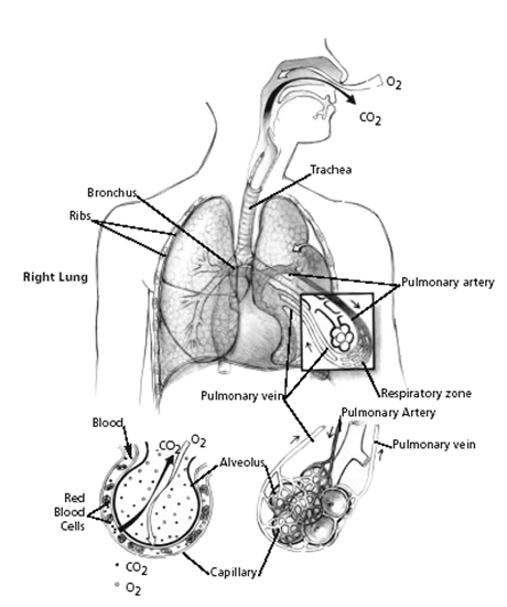
Figure 2: Mechanism of gas exchange.
How respiration is impaired
Many diseases can affect breathing. The most useful classification of respiratory disorders is based on the manner in which the ability of air to come into contact with the hemoglobin in red blood cells (RBCs) is disrupted.
There are only two ways in which respiration can be impaired, regardless of the exact nature of the disorder:
- Disease that prevents adequate amounts of air from reaching the gas-exchange level of the lungs (obstructive lung disease).
- Disease of the lung tissue itself that reduces gas exchange (restrictive lung disease).
Thus, physicians have found it broadly useful to classify respiratory disorders as obstructive or restrictive, or a combination or the two.
Chronic obstructive lung disease (COPD)
Chronic obstructive pulmonary disease (COPD) is the most common type of lung disease. “Obstruction” refers to the fact that air flow in and out of the lungs is impeded. The three most frequent types of COPD in adults are:
- Emphysema
- Chronic bronchitis
- Asthma
In emphysema, lung tissue itself is destroyed. Damaged lung tissue forms non-functional spaces that trap air, and the lungs expand. The effect of these abnormalities is to obstruct air flow to and from the lungs. Emphysema and chronic bronchitis often occur together usually in people with a history of cigarette smoking.
Chronic bronchitis can also occur from long exposure to chemical fumes associated with a particular occupation. Inflammation of the inner surface of bronchi results from exposure to irritating substances. This inflammation, along with excessive secretion of bronchial mucous glands results in bronchial narrowing. Thus, bronchial tree resistance to air flow increases—there is obstruction of air flow.
Restrictive lung disease
The hallmark of restrictive lung disease is loss of usable lung volume, either due to:
- Disease of the gas exchange part of the lungs (lung parenchyma), or
- Some disorder outside of the lungs (extrapulmonary) that prevents air from adequately ventilating normal lung parenchyma.
Examples of parenchymal restrictive lung diseases
Infections (bacterial, fungal, viral, parasitic). Chronic infections that damage lung tissue, such as severe bronchiectasis or advanced pulmonary TB, could result in some degree of restrictive impairment. Radiation, such as used for treatment of cancer, can damage lungs. Radiation lung damage is less of a problem than in the past, because modern equipment can direct therapeutic radiation in beams precisely delivered to the tumor. To the extent that this is impossible because of the size or location of the tumor, it is possible to have fibrotic lung damage caused by radiation.
Inhalation of damaging substances into the lung. Pneumoconiosis is a non-specific term that refers to lung damage from inhaling small particles of some kind. Examples of pneumoconiosis include that caused by asbestos (asbestosis), coal dust (anthracosis), beryllium (berylliosis) silicon dust (silicosis), aluminum, iron, tin (stannosis) and talc. Inhalation of toxic chemicals—such as acidic fumes—can also damage lung tissue.
Drug side-effects.
Autoimmune diseases. Autoimmune disorders such as sarcoidosis, scleroderma, systemic lupus erythematosus (SLE), rheumatoid arthritis (RA), polymyositis, ankylosing spondylitis, and mixed connective tissue disorders can cause pulmonary disease. In all of these disorders, some kind of immune system dysfunction damages the lungs.
Idiopathic diffuse interstitial pulmonary fibrosis (cryptogenic fibrosing alveolitis). This condition causes progressive scarring of the lungs on a microscopic level. This damage can result in decreased gas exchange capacity of the lungs, increasingly severe hypoxemia (lack of oxygen in the blood) and eventually death from respiratory failure. The rate of progression is highly variable, but median survival is less than 3 years. Some cases remain slowly progressive for a number of years, then are triggered by some unknown event to become rapidly more severe.
Other diseases. Examples of other diseases that can cause pulmonary fibrosis include alveolar proteinosis, acquired immunodeficiency syndrome (AIDS), acute respiratory distress syndrome (ARDS), amyloidosis, bone marrow transplantation, cancer, eosinophilic granuloma, eosinophilic pneumonia, lipoid pneumonia, genetic metabolic diseases caused by enzyme deficiencies (Gaucher’s disease, Niemann-Pick disease), Hermansky-Pudlak syndrome, pulmonary vasculitis, tuberous sclerosis, and neurofibromatosis.
Claimants with both lung and heart disease
The close physiological relationship between the cardiovascular and pulmonary systems means an impairment in one of these systems influences the other. For example, it is very common for a claimant with COPD caused or aggravated by cigarette smoking to also have significant cardiac disease or peripheral vascular disease as separate impairments. Thus, if you have both heart and respiratory impairments, you will have a much worse functional restriction than if you had either alone.
Information about your activities
You should take care to provide your doctor with a clear picture regarding the speed at which you can carry out various activities, particularly as limited by shortness of breath. If you state you can climb two flights of stairs, the information is incomplete: (1) Are the stairs climbed at a normal pace? (2) Do you have to stop and rest on the way up, limited by shortness of breath?
Similarly, it is misleading to say you can walk one block or two blocks or three blocks. If the pace is slow enough, some people with chronic pulmonary disease can walk several miles. But if it’s a slow struggle, it hardly represents any practical functional capacity.
Use of the arms can bring on rapid shortness of breath. So it is important to provide times for completion of daily activities, if possible. If not, comparisons to normal individuals can help define functional limitation. For example, “I can walk a block before I get short of breath, but I can’t keep up with my wife” is a much more revealing statement than “I get short of breath walking.”